An X-Ray is a safe, painless, and quick procedure that should have you in and out of our office in no time. Our team of hospital-based radiologists and support staff at Premier Imaging uses advanced equipment to capture high-quality images.

You've probably had X-Rays taken by your dentist or another medical professional, but have you ever wondered exactly how they work, and whether they're safe?
With X-Rays, our experienced Ottawa-based team uses high-energy electromagnetic radiation to produce pictures of the inside of the body. The image produced allows our radiologists to look at bones and joints so we can detect a range of conditions. X-Rays are also sometimes used to detect problems with internal organs and other soft tissues.
If your doctor suspects you may have a fractured or broken bone, heart problem, lung infection, tumour, or another issue, you may be referred to our team of expert radiologists.
Whatever your condition, our experienced radiologists and support staff are here to answer any questions you may have, and to help you feel comfortable and safe.
Different parts of the body absorb energy produced by X-Rays at different rates. After an X-Ray beam passes through the body, a detector on the other side of the body picks up the X-Rays and converts them to an image.
After your X-Rays are complete, a hospital-based radiologist will review your results and ensure you and your doctor receive them in a timely fashion. We are happy to answer any questions you and your healthcare team may have and to schedule any future imaging appointments.
Here is what you can expect during each phase of the X-Rays process at our clinic in Orléans, Ottawa, in addition to details on what to bring with you to your appointment.
Please arrive 5 minutes prior to your appointment to register. No preparations are needed for X-Rays. However, if you wish to avoid changing your clothes, make sure you do not have any metal or plastic on your clothes.
Make sure there is no jewelry in front of the region that needs to be X-Rayed. (i.e. no necklaces for chest, ribs, thoracic or cervical spine).
Prior to some types of X-Rays, you may be given a liquid called a contrast medium that will outline a specific area of your body and help us see more details in the images that are produced. You may swallow the contrast medium, or receive it as an enema or injection.
The technologist will position your body according to the views that are required. During the X-Rays, you'll need to remain still and sometimes hold your breath to avoid moving and creating a blur on the recorded image.
A safe level of radiation will be passed from the machine through your body, to be recorded as an image on a special plate. An X-Ray test is painless and non-invasive.
The entire procedure should take a few minutes for a simple X-Ray. More complex procedures, such as those that require the use of a contrast medium, may take longer.
If you are bringing a child in for an X-Ray, we may use specific techniques or restraints to ensure he or she stays still. These will prevent us from needing to request a repeat procedure if the child moves during the test, and they will not harm your child.
If you choose to remain in the room during the procedure, you'll likely be asked to don a lead apron to prevent unnecessary exposure to radiation.
After your X-Rays, you'll likely be able to get back to your normal activities. Routine X-Rays typically have no side effects.
However, if you needed to take a contrast medium prior to your X-Ray, drink lots of fluids to help clear it from your body. Contact your doctor if you notice pain, redness, swelling, or other symptoms at the injection site.
Because we're able to save your X-Rays digitally to a computer, they can be seen on-screen within minutes.
Our expert radiologists view and interpret your X-Ray, then send a signed report to your doctor, who will explain the results to you. If your case is an emergency, the results can be sent to your doctor within 48 hours.
If follow-up exams are required, your doctor will be able to provide additional information.
Read the answers to our most frequently asked questions about X-Rays at Premier Imaging.
The amount of radiation your body is exposed to during an X-Ray varies depending on the type of organ or tissue being assessed. Children are more sensitive to radiation than adults.
However, since X-Rays use only very low levels of radiation exposure, the risk of cancer is very low. In Ontario, all imaging facilities must follow strict guidelines and regulations related to radiation exposure.
The state-of-the-art imaging systems at our clinic allow us to capture high-quality images efficiently while reducing radiation exposure as much as possible.
The benefits you and your doctor will receive from the test also far outweigh the risk. You may need to wear a lead apron to protect certain parts of your body.
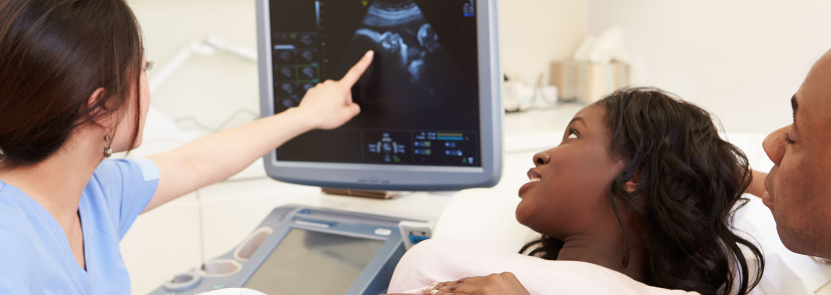
However, if there's a chance you could be pregnant - or if you are pregnant - let your doctor know before having an X-Ray. Though the risk of most diagnostic X-Rays to unborn babies is small, you and your doctor may decide on another imaging test, such as an ultrasound.
In some people, the injection of the contrast medium used to provide better-detailed images can cause side effects such as:
X-Rays help your doctor to diagnose illnesses and health conditions that are not visible during a visual or physical exam.
This test is the quickest and easiest way for your doctor to see and examine injuries, dislocations and fractures of your bones and joints.
After your X-Ray test, no radiation will remain in the body, and there are no side effects from the X-Rays themselves.
Though there is always a slight risk of cancer from excessive radiation exposure, the benefit of an accurate diagnosis far outweighs the risk.
Though the government has defined dose limits for the population and workers who use these machines daily, there is no specific permissible level recommended for patients who require these tests.
Your doctor will compare the medical necessity for an accurate diagnosis to the risk involved. There is no limit for the minimum or maximum number of X-Rays a patient can have within a period of time, or over a lifetime.
X-Ray images provide the most detailed views of bone, but offer little information regarding muscles or tendons.
An MRI may be more useful to help your doctor identify certain types of joint and bone injuries. The MRI can also be used to assess the spinal cord and bone together. Bone bruises or subtle fractures may not visible on X-Ray images.

We'll collaborate with your healthcare team to create a streamlined imaging and diagnostics process. Find out how we can help.