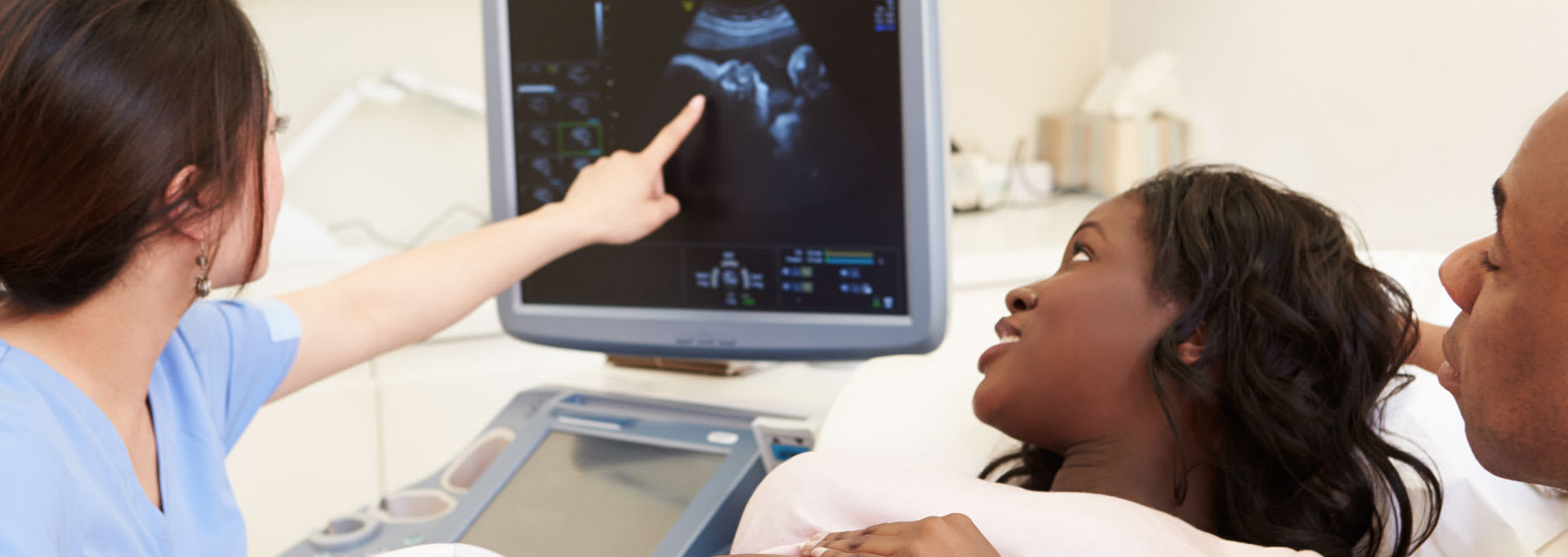
Whether you've had a general ultrasound or not, you may be feeling apprehensive about your upcoming appointment. Our expert radiology team in Ottawa is here to calm your concerns.

Ultrasound testing (sonography) is a safe, useful diagnostic tool that allows us to learn more about the structure and appearance of a patient's internal organs, and to detect any abnormalities that may be in your system.
Depending on the purpose of your ultrasound, you may be feeling joyful and excited (if this is the first time you'll see your baby), or apprehensive (if your doctor suspects one of your body's organs may be causing a health condition). Any or all of these emotions are absolutely normal.
However you may be feeling, and whatever your reason for coming into our ultrasound clinic in Ottawa, our team of expert radiologists and staff are here to support you, answer your questions, help you navigate the process, and keep you calm, cool, and collected.
The non-invasive procedure is painless and uses high-frequency sound waves to produce images in real-time. We are also able to see the blood flowing through blood vessels.
Gel is placed directly on the skin and a transducer (small probe) is used to send the sound waves through the gel into the body. The sounds that bounce back are fed to a computer and transformed into an image.
After your doctor receives the results of your ultrasound, we look forward to addressing any questions or concerns, and guiding you through any follow-up that may be necessary.
Here is what you can expect during each phase of the ultrasound process, along with details on what to bring with you to your appointment at our clinic in Orléans, Ottawa.
Depending on the type of examination you are scheduled to have, preparation instructions may vary.
For some tests, your doctor may tell you not to eat or drink for as many as 12 hours prior to your appointment. Conversely, for other tests you may be instructed to drink up to 6 glasses of water 2 hours before your exam, and to avoid urinating so that your bladder will be full when the test is performed.
Wear clothing that's loose-fitting and comfortable. You may be asked to remove all clothing and jewellery in the area to be examined.
You may also need to wear a gown.
For most ultrasound exams, the radiologist will ask you to lie face-up on an exam table that can be moved or tilted.
During the ultrasound exam, the transducer sends inaudible, high-frequency sound waves into the body, which strike an object (such as organs or tissues). Echo waves then bounce back through the transducer and are recorded to allow us to learn about the size, shape, and consistency of the organ or tissue.
We will also be able to detect changes in the appearance of vessels, tissues and organs, and to detect abnormal masses such as tumours.
The waves are measured by a computer and displayed on a monitor, where we can view a real-time image. Most ultrasound exams are fast (30 minutes to 1 hour in duration), and painless.
Patients do not usually feel any discomfort from the pressure of the transducer, but if the area being examined is tender, you may feel minor pressure or pain.
Rarely, young children may need to be sedated so they can be held still and good quality images can be captured. Parents should ask about this beforehand and make any preparations necessary as requested.
Once the exam has been completed, the ultrasound gel will be wiped from your skin. Though some may be left behind, it will usually dry quickly without discolouring or staining your clothing.
For most patients, ultrasound tests are fast, painless and easily tolerable.
If you have a transesophageal echocardiogram, a transrectal ultrasound or transvaginal ultrasound - where a probe is attached to the transducer and inserted into a natural opening in your body - you may feel minimal discomfort after the procedure.
Most ultrasounds take about 30 minutes, though more extensive exams may take up to an hour.
When the test is complete, you may be asked to dress and wait while the radiologist reviews the images. You should be able to resume normal activities immediately after your appointment.
A radiologist will analyze the images, then send a signed report to the doctor who requested the test. Your doctor will then share the results with you.
If follow-up tests are needed, your doctor will explain why.
Read the answers to our most frequently asked questions about general ultrasounds at Premier Imaging.
The ultrasound scanner used during your test will be made of a computer console, monitor and attached transducer - a small, hand-held device that looks like a microphone.
Inaudible, high-frequency sound waves are sent through the transducer into the body, which transfers returning echoes back to the computer to be recorded, analyzed and displayed on the screen.
A technologist places a small amount of gel on the area of your body that's being examined before the transducer is used. This gel allows the soundwaves to travel back and forth between your body and the transducer.
The video monitor will then display the image in real-time. Your body's structure and/or tissue is analyzed by the computer before it produces the image based on the loudness (amplitude), pitch (frequency) and time it takes for the ultrasound signal to return to the transducer.
At our ultrasound centre, ultrasound imaging is a non-invasive diagnostic medical procedure that helps physicians to diagnose and treat medical conditions.
It can be used to examine many of the body's internal organs, including the heart and blood vessels, eyes, kidneys, liver, pancreas, bladder and others.
Ultrasound can also be used to diagnose heart conditions, guide procedures such as needle biopsies, and guide biopsy of breast cancer.
Benefits
Because air or gas can disrupt ultrasound waves, this test would not be ideal for organs blocked by the bowel, or for a bowel filled with air.
Though ultrasound may be used to detect fluid around or within the lungs, it's not as useful for imaging air-filled lungs.
Similarly, while ultrasound may be used for assessing infection surrounding a bone or for imaging bone fractures, it cannot penetrate bone.
Because sound waves weaken as they pass deeper into the body, large patients are more difficult to image as they have greater amounts of tissue.
You will be instructed not to eat or drink anything for as many as 12 hours before your appointment. If you do eat any food or drink beverages, the ducts and gallbladder will empty so food can be digested and we won't be able to view them easily during the test.
If your appointment is in the morning, ask your doctor when you should eat last the night before. You may take medications with small sips of water.

We'll collaborate with your healthcare team to create a streamlined imaging and diagnostics process. Find out how we can help.