Musculoskeletal pain or injuries can severely impact your quality of life. Our radiologists in Ottawa use MSK ultrasounds to determine the extent of an injury.

An ultrasound technologist performs an MSK (musculoskeletal) ultrasound to specifically focus on your joints and muscles.
Patients often come to us after seeing a doctor about an injury or pain, which may be acute with a rapid onset of severe symptoms, or more chronic and long-lasting. From carpal tunnel syndrome to fibromyalgia, back pain, joint pain, muscle spasms, lumps, and more, musculoskeletal pain can encompass a broad set of conditions.
At Premier Imaging, we understand pain can severely impact your quality of life. By taking an MSK ultrasound, we'll be well on our way to diagnosing your issue and working with your healthcare team to find solutions to help alleviate or manage your pain.
The exam will allow us to determine the extent of the injury or problem, precisely where the source of painful symptoms is located, if it is solid or filled with fluid, and more.
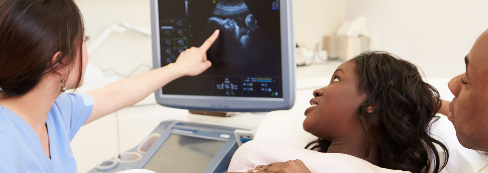
During the ultrasound, a transducer is pressed to the skin and sends high-pitched sound waves to travel through the body. Wave activity is converted into detailed, real-time pictures of tendons, joints, muscles, and other structures.
An MSK ultrasound can help us confirm a diagnosis and follow up with your healthcare team to plan more tests or follow-up diagnostic care.
Here is what you can expect during each phase of the process, along with details on what to bring with you to your appointment at our clinic.
Please wear loose-fitting, comfortable clothing to your appointment, and preferably no jewellery as you may need to remove jewellery and clothing in the area to be examined.
You may also be asked to wear a gown for the procedure.
If a child is the one having the ultrasound, crying or activity can prolong the test as the equipment is very sensitive to motion. Parents or guardians may want to explain the procedure and what will happen to the child, before coming in for the exam.
Also consider bringing small games, toys, music or books to help the time pass.
For most patients, MSK ultrasounds are fast, painless and easily tolerable. These specialized exams focus specifically on a patient's joints and muscles to help determine the extent of an injury. The test is usually done within 15 to 30 minutes.
During the test, you may be asked to sit on a swivel chair or exam table and move the extremity being to be examined, or the radiologist may move it for you so the function of the tendon, joint, ligament or muscle can be assessed.
You may then be asked to lie on the examination table before a warm, clear water-based gel is applied to the area being examined. This gel allows the transducer to connect with the body, and removes air pockets that may form between the skin and transducer. These pockets can block sound waves from entering your body. The transducer is then moved back and forth over the area being examined until the necessary images are produced.
Though you will typically not feel any discomfort during the exam, you may feel pressure or minimal pain if the transducer is passed over a tender area.
The gel will be removed from your skin once the exam is complete, but any gel that is left over will quickly dry and would usually not discolour or stain clothing.
After the exam is complete, you may be asked to dress and wait while the radiologist reviews and analyzes the ultrasound images. After your exam, you should be able to immediately return to your normal activities.
In some circumstances, the radiologist may brief you on the results of your test.
The radiologist will send the results of your exam to the referring doctor, who will then share the results with you. Your doctor will let you know if any follow-up exams are required to determine if there has been any change in abnormalities that were discovered, or if treatment is working or your condition has changed.
Read the answers to our most frequently asked questions about MSK ultrasounds at Premier Imaging.
Muscle aches or arthritis can afflict many people, and anyone experiencing these may suffer from acute or chronic pain, or other symptoms such as inflammation.
Those circumstances may bring you to see us. We also perform MSK ultrasounds for patients with:
Pregnant women are also eligible for this procedure, as musculoskeletal ultrasounds pose minimal risk or discomfort.
Some patients may require surgery and this test may help us determine whether surgical procedures are needed. Later, your doctor may order an MSK ultrasound to assess how well your injury is healing.
With an MSK ultrasound, our trained technologist can capture a clear picture of your tendons, muscles nerves and other internal structures that may be causing pain or inflammation due to strains, tears or arthritis.
Your radiologist can then interpret the ultrasound and share information with your physician, who may be able to gain a better understanding of the source of your specific issue revealed by the data collected.
Ultrasound also doesn't require radiation or the injection of a contrast agent. Plus, you won't feel claustrophobia from being confined to an enclosed space, as you might when undergoing an MRI.
Imaging and healthcare professionals use MSK ultrasounds where appropriate as the procedure is safe, low-cost and fast. We can use Doppler imaging to evaluate for active inflammation where increased blood flow is detected.
This type of ultrasound allows for real-time visualization of the needle and its target during procedures. It can be used for specialized injections into soft tissues and joints, as well as taking samples of fluid (aspiration) from these areas for testing.
With therapeutic injections, it's imperative that medication be injected into precise locations, correctly. Ultrasound allows us to ensure pinpoint accuracy.
If your physician or other members of your health team are able to diagnose your issue after the MSK ultrasound is performed, they may prescribe medicine, physical therapy or other treatments depending on the condition causing your symptoms.
Because ultrasound cannot penetrate bone, we can only use it to view outer surfaces of bony structures - not what is within them (infants are an exception to this rule as they have more cartilage than older children or adults).
This is why we would typically use MRI or other imaging tools to capture images of internal structure of certain joints or bones. We would also not typically use ultrasound to detect causes of back pain.
In addition, larger patients may not be as easy to image as sound waves may not penetrate as deeply as needed to capture high-quality images.

We'll collaborate with your healthcare team to create a streamlined imaging and diagnostics process. Find out how we can help.