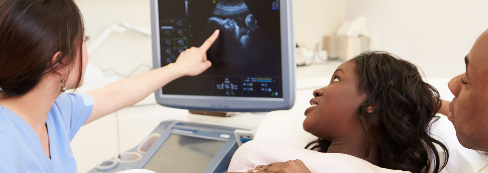
Our expert radiology team in Ottawa uses abdominal ultrasounds to help determine the cause of various conditions, such as stomach pain and kidney stones.

An abdominal ultrasound is a non-invasive procedure used to assess the organs and structures in the abdomen. It uses a transducer that sends out ultrasound waves at a frequency too high to be heard. These are converted into an image of the organs or tissues being examined.
Abdominal ultrasounds can help determine the cause of stomach pain, bloating, kidney stones, liver disease, tumors, and many other conditions. Your doctor may recommend this test if you're at risk of an abdominal aortic aneurysm.
A Doppler ultrasound may also be part of your abdominal ultrasound procedure. A Doppler ultrasound is a special technique used to measure the movement of materials in the body, such as blood flow through arteries and veins.
The sonographer will position the patient on an exam table and apply a water-based gel to the stomach. This gel helps the transducer make secure contact with the body and eliminates air pockets between the transducer and the skin.
The sonographer uses the transducer to assess and capture images of the abdominal organs.
There is no special care required after an abdominal ultrasound. You may resume your usual diet and activities unless your doctor advises you differently.
Here is what you can expect during each phase of the abdominal ultrasound process, along with details on what to bring with you to your appointment at our clinic in Orléans, Ottawa.
Please wear loose-fitting, comfortable clothing to your appointment and preferably no jewelry as you may need to remove jewelry and clothing in the area to be examined.
You may also be asked to wear a gown for the procedure.
If the abdominal structures are being examined, it may be necessary to fast before the exam. If the urinary tract or pelvic organs are being examined, it may be necessary to fill the bladder.
You lie on your back on an examination table for an abdominal ultrasound. A trained professional (a sonographer or radiologist) applies a special gel to your abdomen. The gel works with the ultrasound device to improve image quality.
The device is gently pressed against the stomach by the provider, who moves it back and forth. The device transmits data to a computer. The computer generates images that depict the structures of the abdomen.
An abdominal ultrasound exam usually lasts about 30 minutes.
The gel will be removed from your skin once the exam is complete, but any gel that is left over will quickly dry and would usually not discolour or stain clothing. You should be able to return to regular activities after an abdominal ultrasound.
The images will be analyzed and interpreted by a radiologist. The radiologist will provide the doctor who requested the exam with a signed report. Your doctor will then discuss the findings with you. In some cases, after the exam, the radiologist may discuss the results with you.
A follow-up exam may be necessary to further evaluate a potential problem with additional views or a special imaging technique. It may also check to see if an issue has changed over time. Follow-up exams are frequently the best way to determine whether treatment is effective or whether a problem requires attention.
Read the answers to our most frequently asked questions about abdominal ultrasounds at Premier Imaging.
Abdominal ultrasound can be used to determine the size and location of organs and structures in the abdomen. It can also be used to examine the abdomen for conditions like:
An aortic aneurysm can be detected by measuring the size of the abdominal aorta with ultrasound. Ultrasound can detect stones in the gallbladder, kidneys, and ureters.
Abdominal ultrasound can also help with the placement of needles used to biopsy abdominal tissue or drain fluid from a cyst or abscess.
The benefits of an abdominal ultrasound include:
One treatment advantage of a stomach ultrasound is its ability to screen for abdominal aortic aneurysms.
Screening entails searching for the condition in people who do not have any symptoms. Early detection allows you and your doctor to take action to manage and treat the aneurysm. If an aortic aneurysm ruptures, the ensuing bleeding can be fatal.
For men aged 65 to 75 who have smoked at least 100 cigarettes in their lifetime, a one-time abdominal aortic ultrasound screening is recommended.
Screening is also recommended for men aged 60 and older who have had a parent or sibling with an aortic aneurysm.
If your doctor or other members of your healthcare team are able to diagnose your problem following an abdominal ultrasound, they may prescribe medication or other treatments depending on the condition causing your symptoms.
Air or gas disrupts ultrasound waves. As a result, an abdominal ultrasound scan is ineffective for imaging the air-filled bowel or organs obscured by the bowel.
Although ultrasound is ineffective for imaging air-filled lungs, it can be used to detect fluid around or within the lungs. Similarly, ultrasound cannot penetrate bone, but it can be used to image infections around a bone.

We'll collaborate with your healthcare team to create a streamlined imaging and diagnostics process. Find out how we can help.